We may not have the course you’re looking for. If you enquire or give us a call on +27 800 780004 and speak to our training experts, we may still be able to help with your training requirements.
Training Outcomes Within Your Budget!
We ensure quality, budget-alignment, and timely delivery by our expert instructors.
Mastication means breaking down food into smaller pieces. It is a critical step in the digestive process. To enable this process, the Muscles of Mastication play an important role by controlling the movement of the Mandible (lower jaw).
They also contribute to other vital functions like swallowing and speech. Thus, people beginning their careers in the field of Human Physiology and Biology must learn about these Muscles to dive deeper into the intricacies of the human body. In this blog, you will learn about these Muscles of Mastication and their essential role in Human Physiology.
Table of Contents
1) What are Muscles of Mastication?
2) The primary Muscles of Mastication
a) Masseter muscle
b) Temporalis muscle
c) Lateral pterygoid muscle
d) Medial pterygoid muscle
3) The accessory Muscles of Mastication
4) Conclusion
What are Muscles of Mastication?
The Muscles of Mastication is a group of four powerful Muscles. They work together to control the movement of the Mandible (lower jaw) during chewing. These muscles are basically responsible for all the complex grinding and movements that break down food into smaller and more manageable pieces. This process makes it easier to swallow and digest.
These Muscles are important for proper oral function. It plays a crucial role in the digestive process. They are always in sync, each of them contributing to specific aspects of chewing motion. The precise interplay of these Muscles enables food's complex and efficient breakdown.
The Primary Muscles of Mastication
The following are some of the major Muscles of Mastication that help the digestive process:
a) Masseter Muscle
The Masseter Muscle is a powerful, thick, rectangular muscle. It is located on the sides of the face. It elevates the Mandible (lower jaw) and provides the main force for closing the jaw. It is one of the four Muscles of Mastication, the one responsible for chewing.
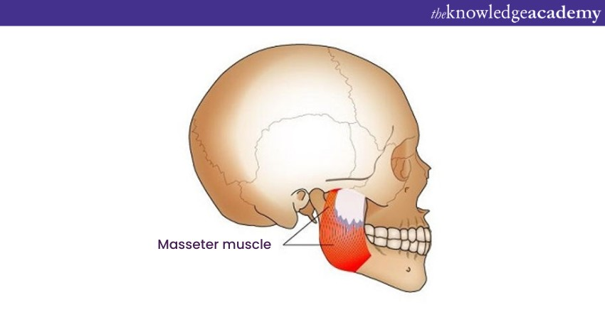
The Masseter Muscle originates from the Zygomatic Arch. It is the bony ridge below the eye. It inserts into the angle and lateral surface of the Mandibular Ramus (the vertical part of the lower jawbone). It consists of two distinct parts:
1) Superficial part
2) Deep part
The masseter muscle plays a crucial role in chewing by elevating the Mandible and providing the primary force for closing the jaw. It works in conjunction with the other Muscles of Mastication to produce the complex grinding and crushing movements that break down food into smaller pieces.
The masseter muscle is innervated by the Masseteric nerve. This nerve is a branch of the Trigeminal nerve's Mandibular division, also denoted as Cranial nerve V3. This nerve transfers signals from the brain to the muscle, controlling its contractions and relaxation. Any condition that affects the masseter muscle can impair chewing ability and lead to temporomandibular joint (TMJ) disorders. Common issues include the following:
a) Masseter muscle hypertrophy: Excessive growth of the masseter muscle can cause a square-shaped face appearance and contribute to TMJ pain and dysfunction.
b) Masseter muscle atrophy: Wasting or shrinking the masseter muscle can weaken the jaw-closing ability and lead to difficulty chewing.
c) Masseter muscle spasm: Involuntary masseter muscle contraction can cause pain, lockjaw, and difficulty opening the mouth.
Ready to improve your lifestyle? Explore our Healthy Lifestyles Training and unlock your path to wellness!
b) Temporalis Muscle
The temporalis muscle is a large, fan-shaped muscle found on the sides of the skull. It extends from the temporal bone, located on eitherside of the head. It is one of the four Muscles of Mastication, responsible for chewing.
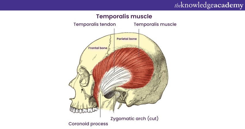
The temporalis muscle originates from the Temporal Fascia, a thin connective tissue covering the temporal bone. It is composed of two main parts that are as follows:
1) Superior part
2) Inferior part
The temporalis muscle plays a significant role in chewing by elevating the Mandible and contributing to grinding and crushing movements. It works in synergy with the other Muscles of Mastication to break down food into smaller pieces.
The temporalis muscle is innervated by the temporal branches of the Mandibular division of the Trigeminal nerve (cranial nerve V3). These nerve branches transmit signals from the brain to the muscle, controlling its contraction and relaxation. Any condition that affects the temporalis muscle can impair chewing ability and lead to Temporomandibular Joint (TMJ) disorders. Common issues include the following:
a) Temporalis muscle hypertrophy: Excessive growth of the temporalis muscle can cause a bulging or fullness on the sides of the head and contribute to TMJ pain and dysfunction.
b) Temporalis muscle atrophy: Wasting or shrinking the temporalis muscle can weaken the jaw-closing ability and lead to difficulty chewing.
c) Temporalis muscle spasm: Involuntary contraction of the temporalis muscle can cause pain, headaches, and difficulty opening the mouth.
c) Lateral pterygoid muscle
The lateral pterygoid muscle is a two-headed muscle. It is located deep within the Infratemporal Fossa, a complex area located at the base of the skull. It is also one of the four Muscles of Mastication.
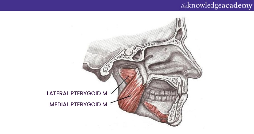
The lateral pterygoid muscle consists of two distinct heads, that are as follows:
1) Superior head
2) Inferior head
The lateral pterygoid muscle plays a versatile role in chewing, contributing to various mandibular movements. Its primary functions include the following:
a) Protrusion: It protracts the Mandible, moving it forward and allowing incisor biting.
b) Lateral excursion: It moves the Mandible laterally (side-to-side), enabling grinding and crushing motions.
c) Depression: It assists in depressing the Mandible, contributing to jaw opening.
The lateral pterygoid muscle works simultaneously with the other Muscles of Mastication to produce the complex movements required for effective chewing. The Lateral Pterygoid nerve innervates the lateral pterygoid muscle. It is a branch of the Trigeminal nerve's Mandibular division, also called the Cranial nerve V3. This nerve sends signals from the brain to the muscle, controlling its contraction and relaxation. Common issues include the following:
1) Lateral pterygoid muscle spasm: Involuntary muscle contraction can cause pain, difficulty opening the mouth, and a clicking or popping sensation in the TMJ.
2) Lateral pterygoid muscle injury: Damage to the muscle can result from trauma, overuse, or inflammation, leading to pain, chewing difficulties, and TMJ dysfunction.
3) Temporomandibular disorders: TMJ conditions can involve the lateral pterygoid muscle, contributing to pain, clicking or popping sounds, and limitations in jaw movement.
Do you want to gain in-depth knowledge about Nutrition to improve your physical well-being? Sign up now for our Nutrition Training!
d) Medial Pterygoid Muscle
The medial pterygoid muscle is a thick, strap-like muscle located deep within the Infratemporal Fossa of the skull.
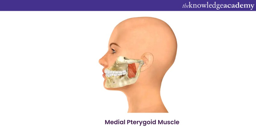
The medial pterygoid muscle originates from the Medial Pterygoid plate (a bony projection of the sphenoid bone) and inserts into the Angle of the Mandible (lower jaw). It is composed of two distinct parts, namely:
1) Superficial part
2) Deep Part
The medial pterygoid muscle is innervated by the Medial Pterygoid nerve, a branch of the Mandibular division of the Trigeminal nerve. It is also referred to as the Cranial nerve V3. This nerve transmits signals from the brain to the muscle, controlling its contraction and relaxation.
Any condition that affects the medial pterygoid muscle can impair chewing ability and lead to temporomandibular joint (TMJ) disorders. Common issues include the following:
a) Medial pterygoid muscle hypertrophy: Excessive growth of the medial pterygoid muscle can cause a bulge or fullness on the sides of the face and contribute to TMJ pain and dysfunction.
b) Medial pterygoid muscle atrophy: Wasting or shrinking of the medial pterygoid muscle can weaken the jaw-closing ability and lead to difficulty chewing.
c) Medial pterygoid muscle spasm: Involuntary contraction of the medial pterygoid muscle can cause pain, difficulty opening the mouth, and a clicking or popping sensation in the TMJ.
Improve your Nutrition and health with our Nutrition and Fitness Training. Sign up now!
The Accessory Muscles of Mastication
There are some more muscles which help in the Mastication process. These muscles are listed below:
a) Buccinator
The buccinator muscle is a thin, quadrilateral muscle that forms the lateral wall of the oral cavity and the cheek. The buccinator muscle originates from the upper jawbone and the Ramus of the lower jawbone. It further inserts into the Modiolus, a structure at the corner of the mouth. It plays a variety of roles in facial movements and chewing. Here's how:
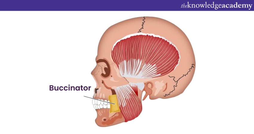
The buccinator muscle originates from the Maxilla (upper jawbone) and the Ramus of the Mandible (lower jawbone). It further inserts into the Modiolus, a fibrous structure at the corner of the mouth. It plays a variety of roles in facial movements and chewing. Here's how:
1) Chewing
2) Facial expressions
3) Swallowing
The buccinator muscle is stimulated by the Buccal nerve. Division of the Trigeminal nerve or Cranial nervethis nerve is a branch of the Mandibular V3. This nerve also transmits signals from the brain to the muscle, controlling its contraction and relaxation. Any condition that affects the buccinator muscle can impair facial movements, chewing ability, and swallowing. Common issues include the following:
a) Buccinator muscle paralysis: Damage to the buccal nerve can paralyse the buccinator muscle. This can lead to facial asymmetry, difficulty chewing, and drooling.
b) Buccinator muscle spasm: Involuntary contraction of the buccinator muscle can cause pain and difficulty opening the mouth.
c) Buccinator muscle hypertrophy: Excessive growth of the buccinator muscle can cause a square-shaped face appearance.
d) Buccinator muscle atrophy: Wasting or shrinking of the buccinator muscle can lead to difficulty chewing and facial sagging.
b) Suprahyoid muscles
The suprahyoid muscles are a group of four muscles located above the hyoid bone in the neck. They are responsible for elevating the Hyoid bone involved in swallowing and speech. The four suprahyoid muscles include the following:
1) Digastric muscle: It is a two-bellied muscle that begins from the Mastoid process of the Temporal bone and inserts into the digastric fossa of the Mandible. It elevates the hyoid bone and helps to open the mouth.
2) The stylohyoid muscle: It originates from the Temporal bone's Styloid process and ends in the greater horn of the Hyoid bone. It elevates and retracts the hyoid bone
3) The geniohyoid muscle: The geniohyoid muscle originates from the mental spine of the Mandible and ends in the body of the Hyoid bone. It elevates and protrudes the Hyoid bone.
4) Mylohyoid muscle: It is a broad, fan-shaped muscle that forms the floor of the mouth. It originates from the mylohyoid line of the Mandible and inserts into the Raphe Pterygomandibulare. It elevates and tenses the floor of the mouth.
5) The suprahyoid muscles: They work together to elevate the hyoid bone, which is necessary for swallowing and speech. They also help to open the mouth and tense the floor of the mouth.
c) Infrahyoid Muscles
The infrahyoid or strap muscles are a group of four paired muscles below the Hyoid bone in the neck. They are responsible for depressing the Hyoid bone during swallowing and stabilising the larynx during speech. The four infrahyoid muscles are as follows:
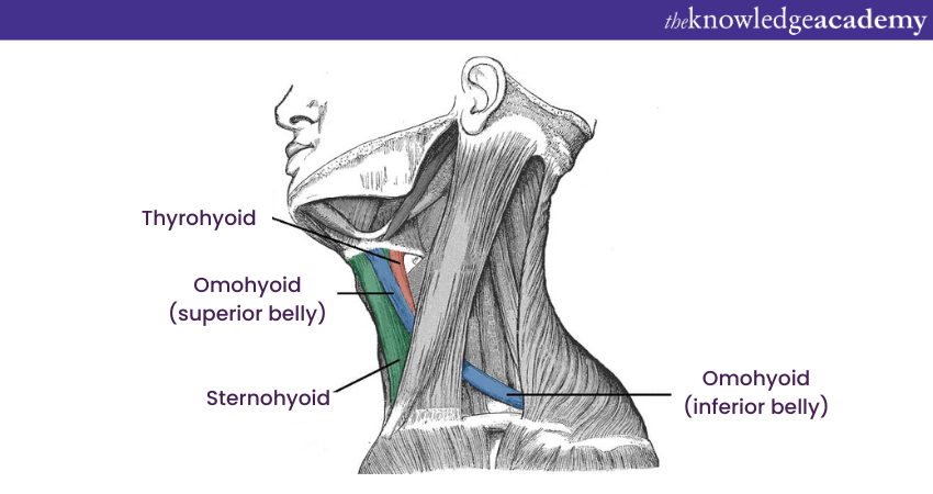
1) The sternohyoid muscle: Originates from the Sternum (breastbone) and inserts into the Hyoid bone. It depresses the Hyoid bone.
2) The Sternothyroid muscle: Originates from the Sternum (breastbone) and ends in the Thyroid Cartilage of the Larynx. It depresses the Larynx.
3) The Thyrohyoid muscle: Originates from the Larynx's Thyroid Cartilage and inserts into the Hyoid bone. It depresses the Hyoid bone and elevates the larynx.
4) The Omohyoid muscle: It is a long, thin muscle that actually originates from the Scapula (shoulder blade), passes over the clavicle (collarbone), and inserts into the hyoid bone. It helps to stabilise the larynx and depress the hyoid bone.
5) The Infrahyoid muscles: They work together to depress the hyoid bone, which is necessary for swallowing. They also help to stabilise the larynx during speech.
Are you eager to learn about essential life lessons? Sign up now for our Life Coach Training!
Conclusion
After reading this blog, we hope you are clear about the different Muscles in Mastication. These Muscles are an essential part of human physiology. They help digestion by breaking down complex foods into smaller pieces, making it easy for you to swallow and digest.
Learn how to build a healthy life with our Active and Healthy Lifestyles Training. Join now!
Frequently Asked Questions

The muscles of mastication are supplied by the mandibular branch of the trigeminal nerve (cranial nerve V3). This nerve innervates the masseter, temporalis, medial pterygoid, and lateral pterygoid muscles. These muscles enable the jaw movements required for chewing.

The strongest muscle of mastication is the masseter. It is capable of exerting significant force, enabling powerful jaw movements necessary for chewing.

The Knowledge Academy takes global learning to new heights, offering over 30,000 online courses across 490+ locations in 220 countries. This expansive reach ensures accessibility and convenience for learners worldwide.
Alongside our diverse Online Course Catalogue, encompassing 17 major categories, we go the extra mile by providing a plethora of free educational Online Resources like News updates, Blogs, videos, webinars, and interview questions. Tailoring learning experiences further, professionals can maximise value with customisable Course Bundles of TKA.

The Knowledge Academy’s Knowledge Pass, a prepaid voucher, adds another layer of flexibility, allowing course bookings over a 12-month period. Join us on a journey where education knows no bounds.

The Knowledge Academy offers various Healthy Lifestyles Training, including Active and Healthy Lifestyles Training, Cognitive Behavioural Therapy Training, and Yoga Training. These courses cater to different skill levels, providing comprehensive insights into Mindfulness.
Our Healthy Lifestyle Blogs cover a range of topics related to Earned Value Management, offering valuable resources, best practices, and industry insights. Whether you are a beginner or looking to advance your Health and Safety Skills, The Knowledge Academy's diverse courses and informative blogs have got you covered.
Upcoming Health & Safety Resources Batches & Dates
Date
 Life Coach Training
Fri 14th Feb 2025
Fri 11th Apr 2025
Fri 13th Jun 2025
Fri 15th Aug 2025
Fri 10th Oct 2025
Fri 12th Dec 2025


 Top Rated Course


 If you wish to make any changes to your course, please


